PARALLEL TWIN QUARTZ SPLINT FRC FOR NATURAL TOOTH PONTIC
04/14/2023
The first response to an injury or eventual infection developing in a tooth is by the body’s defense mechanism itself, which initiates inflammatory response in that area in order to heal it. The second response is by the patient himself who seeks dental treatment in order to eliminate pain and discomfort. The third response is by the Dentist by means of intervention thereby employing well established treatment protocols such as diagnostic procedures to know tooth vitality status, investigations such as X-Ray etc. and initiate treatment such as Root Canal treatment and medications. Failing which second line of treatment is indicated such as more medicaments, higher investigations such as CBCT and more invasive procedures such as Apicoectomy/Root Re-section etc. If all of the above fail to save the tooth then extraction is suggested. After having undergone so much in terms of failed efforts to save the tooth and having to come to the point of loosing that very tooth the patient is usually demotivated to undergo future surgeries such as Dental implant Procedures. Neither is the patient willing to wear a RPD nor undergo tooth reduction for an FPD. What if the same tooth crown is placed in its natural position permanently with Fiber Reinforced Composite materials. That’s a treatment plan that the patient is very willing to accept, keeping in mind its benefits of being:
1. Non-invasive
2. Quick
3. Much less expensive compared to an implant or an FPD
4. Fixed replacement
5. Esthetically as good as it gets
6. Supportive to healing processes of the body
7. Easy to maintain for hygiene accessibility and repairable by dentist if required.
Here I present a case of a Natural Tooth Pontic (NTP) Maxillary Lateral incisor restoration by Fiber Reinforced Composite FRC Material. The FRC material I used here is QUARTZ SPLINT™ by RTD France. The supporting materials used are EXACLEAR, G-PREMIO BOND and GAENIAL ANTERIOR composite by GC Corporation Japan. The step-by step easy to follow procedure is given with each photograph.
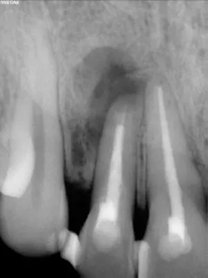
FIG 1:
X-Ray of 36 year old malepatient who had undergone repeated root canal treatment, two apicoectomy procedures and a poorly done stabilisation over the past few years at some other clinic. But there was a fistula with purulent discharge and accompanying pain and inflammation in the region warranting tooth extraction.
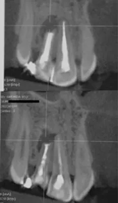
FIG 2:
CBCT of the patient indicates to an unsalvageable tooth.
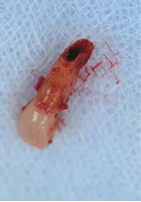
FIG 3:
This sectional image from the CBCT shows the extent of bone damage caused by the lesion.
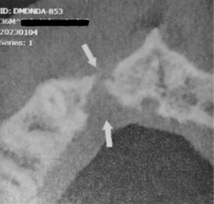
FIG 4:
The extracted tooth clearly shows lack of retrograde filling.
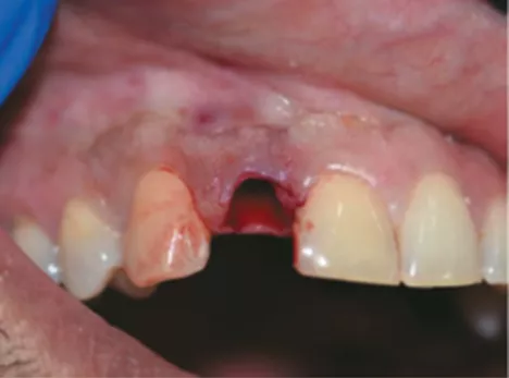
FIG 5:
The extraction socket just after extraction of tooth #12. There was no buccal cortical bone. Curettage was performed to remove perapical cyst and granulation tissue. PRF grafting was done for better tissue healing and one week’s waiting time was observed post extraction.
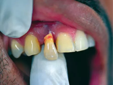
FIG 6:
The extracted lateral incisor shown here in the process of being cut and contoured to size to be used as a Natural Tooth Pontic which will be cleaned and filled from the cervical/apical area with composite resin.
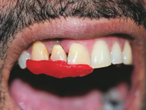
FIG 7:
GC Pattern Resin was used to position the tooth in its correct location after preparing the Pontic as desired and functional occlusal considerations.
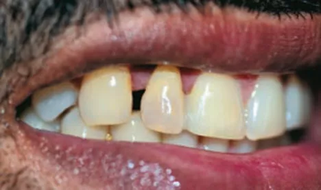
FIG 8:
This shows the initial stabilisation of the NTP being bonded to the central incisor with GC’s GPremio Bond which is a Universal bond along with GC’s Gaenial Injectable Composite after total etch technique. Now the tooth is in its favourable position
and will be strengthened with FRC material.
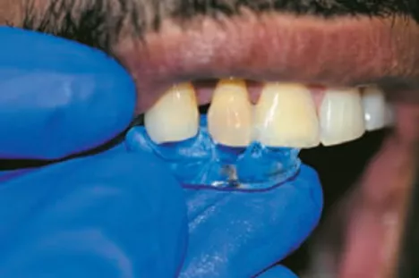
FIG 9:
Palatal side clear A-Silicon Index was made using GC’s Exaclear material to adapt FRC closely to the tooth surface. A small channel was prepared on the incisal third area of Central and Lateral incisor teeth to receive the FRC material being adapted with this index through which initial light curing was done. The high transparency of Exaclear makes it possible to do so.
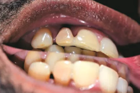
FIG 10:
This incisal view shows reinforcement of the bond between Centre and Lateral incisor on the palatal side now joined with FRC material and covered on top with composite material
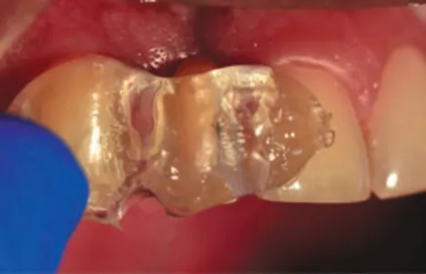
FIG 11:
Exaclear (GC) Index was made now for the labial side adaptation of the FRC
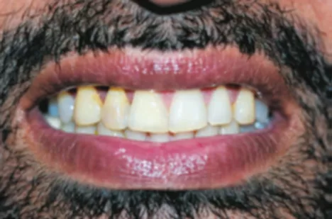
FIG 12:
Tooth #12 and #11 now joined together which give a natural appearance but further strengthening is needed from a bio mechanical perspective for longevity of the restoration
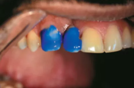
FIG 13:
Etching is done for one minute prior to doing the rinsing, drying, bonding, material application and light curing steps.
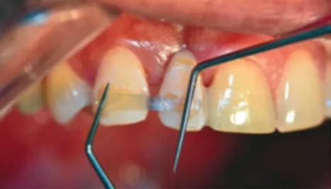
FIG 14:
A channel was prepared in the enamel of teeth #13 and #12 to receive QUARTZ SPLINT (RTD France) which is composed of quartz fibres pre-wetted with a light curable bonding resin. Here Quartz Splint UD was used which comprises of unidirectional quartz fibres. Initial adaptation was done with the help of fine explorers as shown, the FRC material was held in place with clear Index as shown in Figure 11. And light cured in place along with bonding agent.
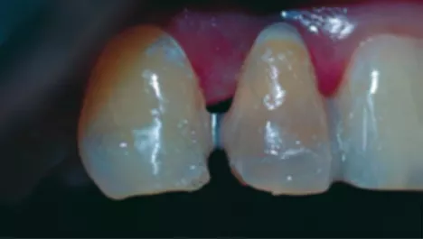
FIG 15:
An additional piece of Quartz Splint UD is now added for enhanced structural strength. It will also be adapted and cured using the Exaclear Index
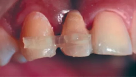
FIG 16:
This photo shows almost invisible optical properties of Quartz Splint (RTD) and Gaenial Injectable (GC) when bonded together using G-Premio Bond (GC). Now the NTP lateral incisor is FRC bonded from the labial side to the canine and from the palatal side to the Central incisor. In essence, sandwiched between PARALLEL TWIN QUARTZ SPLINT FRC.
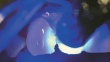
FIG 17:
While the initial light curing to adapt the FRC is done through the clear index, each time additional and thorough light curing for at least 30 seconds is done after removal of the index so that the pre- soaked FRC and the covering resin (Gaenial Anterior, GC in this case) is fully cured.
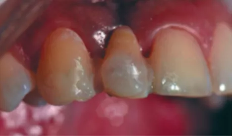
FIG 18:
Now that the FRC adapted and covering resin along with FRC are fully cured, additional injectable composite is used to strengthen inter dental incisal/middle tooth area between #13 and #12
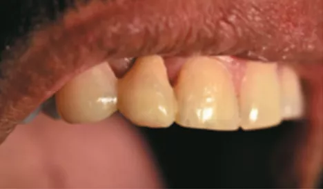
FIG 19:
The areas of Quartz Splint (RTD) and partially veneered surface of the teeth covered with Gaenial Anterior (GC) are finished and polished.
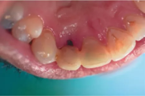
FIG 20:
FRC Natural Tooth Pontic (NTP) as seen from the palatal side. The soft tissue remodelling and regional maturation of the tissue will continue in a natural way on the palatal side now that the infective lesion has been treated. The pontic has been shaped like a modified ridge lap pontic to allow self cleansing of the area. If there is much gap formation due to remodelling of the soft tissue underneath the NTP after few years, it can be filled without disturbing the prostheses using regular etching and bonding composite.
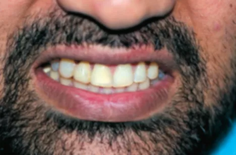
FIG 21:
The patient’s smile is restored in the most conservative, least traumatic and in a naturally aesthetic manner in minimum time and maximum economic savings to the patient.
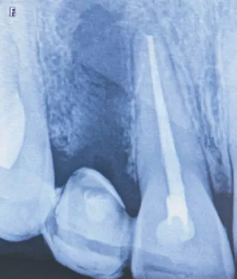
FIG 22:
The X-Ray of the patient after restoration

Dr. Mahesh Chauhan has more than a 100 lectures/ publications to his credit. His name has featured in Marquis Who’s Who in Healthcare and Medicine 2006, 6th edition, published from USA. He is the author of the book titled : A Visual Guide to Fiber Reinforced Composites in Dentistry by Wiley India Pvt. Ltd.
Source: Article Citation Chauhan, M. (2023). Parallel twin quartz splint FRC for natural tooth pontic. Dental Practice, 19(1), 28-31

